
When your doctor orders a brain MRI, it’s not because they’re being overly cautious-it’s because they need to see what’s happening inside your brain with unmatched clarity. Unlike X-rays or CT scans, MRI doesn’t use radiation. Instead, it uses powerful magnets and radio waves to create detailed pictures of your brain’s soft tissues. This makes it the best tool for spotting things like small tumors, early strokes, inflammation, or the subtle changes of multiple sclerosis-problems that might be invisible on other scans.
What You’re Actually Seeing on a Brain MRI
A brain MRI doesn’t show a single image. It shows multiple layers of information, each captured using a different technical setting called a pulse sequence. These sequences highlight different tissues based on how water molecules behave inside them. Think of it like taking photos of the same scene under different lighting-each one reveals something new.
The most common sequences you’ll see are T1-weighted, T2-weighted, FLAIR, DWI, and SWI. On a T1 image, fat looks bright white, and cerebrospinal fluid (CSF)-the liquid that surrounds your brain-looks dark. This is great for seeing anatomy: where the gray matter ends and the white matter begins, where the ventricles (fluid-filled spaces) sit, and whether any structures are shifted or compressed.
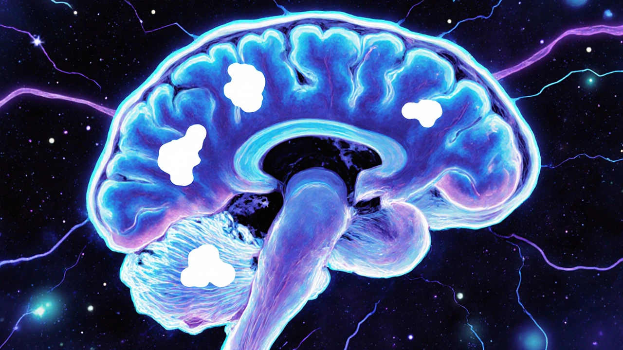
T2-weighted images flip the script. Here, water is bright. That means areas with swelling, infection, or damage-like a stroke or tumor-show up as glowing white spots. But here’s the catch: CSF is also bright on T2. That can make it hard to tell if a bright spot is actual damage or just fluid. That’s where FLAIR comes in.
FLAIR is like a filter that turns off the CSF signal. Now, any bright spots near the ventricles or along the brain’s surface are almost certainly abnormal. This is critical for spotting multiple sclerosis plaques, which often form around the brain’s fluid spaces. Without FLAIR, those lesions could be missed or mistaken for normal fluid.
Then there’s DWI-diffusion-weighted imaging. This sequence is a game-changer for stroke. Within minutes of a stroke starting, water movement in brain cells gets blocked. DWI picks this up immediately. A bright spot on DWI with a dark spot on the matching ADC map? That’s an acute stroke. It’s so reliable that in emergency rooms, a normal DWI MRI can rule out stroke faster than waiting hours for CT changes to appear.
Finally, SWI (susceptibility-weighted imaging) is like a metal detector for blood. It picks up tiny amounts of iron from old bleeds, even ones you never knew about. These microhemorrhages can show up in people with high blood pressure, Alzheimer’s, or after head injuries. Finding them helps doctors understand if a patient’s symptoms are linked to long-term vascular damage.
Common Findings-and What They Really Mean
Not every bright spot on an MRI is a crisis. Many findings are common, especially as people age. Here are the most frequent ones you’ll see, and what they actually indicate.
White matter hyperintensities-small bright spots in the brain’s white matter-are seen in 15% of people under 50 and up to 90% of those over 70. They’re often linked to small blood vessel disease from high blood pressure or aging. On their own, they don’t mean dementia or MS. But if they’re widespread, especially in the deep parts of the brain, they suggest vascular cognitive decline. The key is looking at the pattern: scattered dots? Likely aging. Large patches near the ventricles? Could be MS.
Lacunar infarcts are tiny (3-5mm) strokes in deep brain areas like the thalamus or basal ganglia. They’re often silent-meaning the person never had symptoms like weakness or slurred speech. But they’re a warning sign. Each one means small arteries in the brain are damaged. Multiple lacunes increase stroke risk over time.
Brain atrophy-shrinkage of brain tissue-is normal with aging. But if the hippocampus (memory center) is shrinking faster than expected, or if the ventricles are enlarged beyond what’s typical for age, it could point to early Alzheimer’s or another neurodegenerative condition. The trick is comparing the scan to what’s normal for the person’s age group.
Incidental findings happen more than you think. A small meningioma (usually harmless tumor), a vestibular schwannoma (acoustic neuroma), or even a tiny aneurysm might show up on a scan done for headaches or dizziness. Most are slow-growing and never cause problems. But they need follow-up. A 2mm tumor in the cerebellopontine angle might not need surgery now, but it should be checked again in 6-12 months.
When MRI Is the Right Choice-and When It’s Not
Doctors don’t order brain MRIs lightly. In fact, the American College of Radiology says MRI is usually not appropriate for people with uncomplicated migraines. Why? Because only about 1.3% of scans in people with routine headaches show anything meaningful. That’s why guidelines stress: order MRI only if there are red flags.
Red flags include: new seizures, sudden vision changes, progressive weakness, confusion, balance problems, or headaches that wake you up at night or get worse with bending over. If you’re 50 and have a new type of headache, that’s a red flag. If you’ve had the same headache for 20 years and it’s unchanged? Probably not.
CT scans still have their place. In trauma, when someone’s unstable or needs quick answers, CT wins. It’s faster, cheaper, and better at spotting skull fractures or fresh bleeding. But if the CT is normal and symptoms persist, MRI is the next step. For example, if someone has dizziness and hearing loss, a CT might show nothing. An MRI can reveal a 2mm acoustic neuroma-something CT would miss entirely.
How to Read an MRI Like a Pro (Without Being a Radiologist)
You don’t need to be a doctor to understand the basics. Start with the midline. Is the brain centered? Are the ventricles symmetrical? If one side is swollen or compressed, that’s a problem.
Next, check the cerebellum and brainstem. These areas are hard to see on CT but crystal clear on MRI. A lesion here could explain dizziness, double vision, or trouble walking.
Then, look at the white matter. Are there bright spots? Are they scattered or clustered? Are they near the ventricles? That’s the MS pattern. Are they deep in the brain, near the base? That’s more likely vascular.
Finally, check the meninges-the layers around the brain. Are they thickened? That could mean infection or inflammation. Are there abnormal enhancements after contrast? That suggests active disease like a tumor or abscess.
And always check the cerebellopontine angle. It’s a tiny space behind the ear. Many radiologists miss it. But that’s where small tumors hide. A quick glance here can catch a problem early.
What MRI Can’t Tell You
Even the best MRI has limits. It can’t tell you how long a lesion has been there. A bright spot could be a new stroke-or one from 10 years ago. Without contrast or special sequences, age is guesswork. That’s why doctors look at your symptoms and history. An MRI is a snapshot. It needs context.
It also doesn’t show brain function. You might have a lesion, but if you’re not having symptoms, it might not be causing harm. That’s why a normal MRI doesn’t rule out neurological disease-some conditions, like early Parkinson’s or some types of dementia, don’t show structural changes until later.
And MRI can’t diagnose everything. Conditions like chronic pain syndromes, fibromyalgia, or functional neurological disorder don’t show up on scans. That doesn’t mean they’re not real. It just means the tools we have now aren’t sensitive enough.
What’s New in Brain MRI?
Technology is moving fast. Ultra-high-field 7.0T MRI machines now show brain layers in incredible detail-useful for research and complex epilepsy cases. AI tools are cutting scan times in half without losing quality. Some software can automatically detect MS plaques or measure brain volume changes over time.
Quantitative MRI is the next frontier. Instead of just looking at pictures, doctors are starting to measure things: how fast water moves through white matter (DTI), how much myelin is present, or how much blood flows through brain tissue. These numbers could one day replace guesswork with real data.
But access is uneven. In rural areas, only 42% of hospitals have MRI machines. In cities, it’s 91%. That means people in remote areas often wait longer-or get a less detailed CT instead.
What to Do After Your Scan
Don’t panic if you see a “bright spot” on your report. Most findings are not emergencies. Ask your doctor: Is this new? Is it related to my symptoms? Does it need follow-up? If the radiologist says “likely benign” or “chronic,” that’s usually good news.
If you’re told you have MS, a tumor, or a vascular issue, ask for a copy of the images. Don’t rely on the report alone. Get a second opinion from a neuroradiologist if the findings are unclear or serious.
And remember: an MRI is a tool-not a verdict. It helps guide decisions, but your symptoms, history, and physical exam matter just as much.
Can a brain MRI detect Alzheimer’s disease?
A standard brain MRI can’t diagnose Alzheimer’s directly, but it can show signs that suggest it. The most common finding is shrinkage (atrophy) of the hippocampus and other memory-related areas, especially when it’s more than expected for the person’s age. While MRI can’t see amyloid plaques (the hallmark of Alzheimer’s), it helps rule out other causes like tumors or strokes. Newer techniques like amyloid PET scans or quantitative MRI measuring brain volume changes over time are becoming more common in specialized centers.
Why do I need contrast for some brain MRIs?
Contrast dye (gadolinium) highlights areas where the blood-brain barrier is broken. That happens in active inflammation, tumors, infections, or recent strokes. If a lesion lights up after contrast, it means the problem is currently active. Without contrast, you might see an old scar or a stable tumor-but you won’t know if it’s growing or causing inflammation. Contrast helps doctors decide if treatment is needed now or if it’s safe to wait.
Are brain MRIs safe for everyone?
Most people can have a brain MRI safely. But it’s not for everyone. People with certain implants-like pacemakers, cochlear implants, or old aneurysm clips-may not be able to have one because of the strong magnetic field. Metal fragments in the eyes are also dangerous. If you’ve had surgery, always tell the technologist. Pregnancy is not a strict contraindication, but contrast is usually avoided in the first trimester. Claustrophobia can be an issue too; open MRI machines or sedation can help.
How long does a brain MRI take?
A standard brain MRI takes about 30 to 45 minutes. If contrast is needed, it adds another 10-15 minutes. High-resolution scans for small structures like the inner ear or pituitary gland can take longer. You have to lie still the whole time-even small movements blur the images. If you’re anxious or in pain, ask about sedation options beforehand. Some centers offer headphones with music to help you relax.
Can an MRI show the cause of my headaches?
In most cases, no. If your headaches are typical migraines or tension headaches, your MRI will likely be normal. That’s actually good news-it means there’s no structural cause. MRI is only ordered if there are warning signs: sudden severe headaches, headaches that wake you up, headaches with vision changes, weakness, or confusion. If those are present, MRI can find tumors, aneurysms, or inflammation. But for routine headaches, it’s rarely helpful and often leads to unnecessary worry.
What’s the difference between a 1.5T and 3.0T MRI machine?
The number stands for tesla, the unit of magnetic field strength. A 3.0T machine is twice as strong as a 1.5T. That means better image detail, especially for small structures like cranial nerves, tiny tumors, or early MS plaques. It’s also faster. But 3.0T can exaggerate certain artifacts and isn’t always better for people with metal implants. Most hospitals use 1.5T for routine scans and 3.0T for complex cases. For most neurological conditions, both are accurate-3.0T just gives more detail.
Tom Shepherd
November 27, 2025 AT 22:47I've had two MRIs in the last five years and still don't know what half the terms mean. T1, T2, FLAIR - it's like they're speaking a different language. I just hope the radiologist knows what they're looking at.
Frances Melendez
November 28, 2025 AT 09:20People these days get MRIs for headaches like it's a spa day. No wonder healthcare is broke. If you can't handle a little discomfort or anxiety, maybe you shouldn't be demanding expensive scans. My grandfather had a stroke and didn't even have access to a CT, let alone an MRI. Stop being so entitled.
Jonah Thunderbolt
November 28, 2025 AT 20:11Let me just say - the fact that you’re even discussing ‘white matter hyperintensities’ without mentioning the gut-brain axis and the role of chronic low-grade inflammation mediated by IL-6 and TNF-alpha… is frankly, amateurish. We’re talking about neurodegenerative cascades here, not a PowerPoint slide from med school 2008. And don’t get me started on the lack of discussion about glymphatic clearance - which, FYI, is the real key to understanding those ‘incidental findings.’ 😔
reshmi mahi
November 29, 2025 AT 12:32USA spending billions on 3T machines while India still uses 1990s machines for stroke patients? Of course. Your country buys fancy toys while ours saves lives. But hey, at least you can post about it on Reddit. 🤷♀️
laura lauraa
December 1, 2025 AT 06:20I find it profoundly disturbing that the medical establishment continues to treat brain imaging as diagnostic gold when, in reality, it is merely a static, anatomical snapshot - utterly incapable of capturing the dynamic, phenomenological reality of subjective neurological experience. The reductionism is not just scientific - it is moral. And yet, we continue to allow it to dictate treatment. How many lives have been lost to the illusion of objectivity?
Kaleigh Scroger
December 2, 2025 AT 23:53For anyone scared of what they might see on an MRI - this is the thing nobody tells you. Most things that show up are just background noise. White matter spots? Probably just high blood pressure or aging. Tiny aneurysms? 95% never rupture. Even MS plaques can be silent for years. The scan doesn't define you. Your symptoms do. Your doctor’s context does. Your life does. Don’t let a bunch of grayscale pixels scare you into panic. Ask for the report in plain language. Get a second opinion if it feels off. But don’t Google it at 3am.
Elizabeth Choi
December 4, 2025 AT 16:42Interesting how the article glosses over the fact that 70% of incidental findings lead to unnecessary follow-ups, biopsies, or anxiety-driven visits. The real cost isn’t the machine - it’s the downstream healthcare cascade. We’re overdiagnosing at scale.
Iives Perl
December 6, 2025 AT 06:45MRIs are controlled by the government to track brain activity. That’s why they never show the real cause of migraines - they’re hiding the mind control signals. You think the 3T machine is for tumors? Nah. It’s for detecting dissent. They’re mapping your thoughts. Stay alert.
Savakrit Singh
December 7, 2025 AT 02:47It is imperative to underscore that the utilization of MRI technology in developing nations remains critically under-resourced. The disparity in access, as referenced in the article, is not merely logistical but emblematic of a broader epistemological hegemony wherein Western diagnostic paradigms dominate global neurology. This is a colonial legacy in medical practice.
Jebari Lewis
December 7, 2025 AT 20:44I’ve spent the last year studying neuroimaging literature after my wife’s diagnosis and I want to say thank you for this. You broke it down like a teacher who actually cares. I showed it to our neurologist and he nodded and said ‘you’ve done your homework.’ That means more than any scan. Please keep writing like this. People need clarity, not jargon.
sharicka holloway
December 9, 2025 AT 06:51My dad had a 2mm meningioma found on a scan for dizziness. They said watch and wait. Five years later, still no symptoms. He’s hiking in Colorado now. Don’t let a dot on a screen steal your peace. You’re more than your MRI.
Alex Hess
December 9, 2025 AT 18:02This is just a glorified textbook summary. No original insight. Where’s the critique? Where’s the depth? You’re just regurgitating radiology 101. I expected better.
Leo Adi
December 10, 2025 AT 12:09In India, we say ‘MRI is like a mirror - it shows what’s there, but not why it’s there.’ My uncle had a bright spot. Doctors said ‘age.’ But his memory faded. We found out later it was a slow-growing tumor. Sometimes the mirror lies. Always ask: is this the beginning, or just the end of something already gone?
Melania Rubio Moreno
December 12, 2025 AT 04:36FLAIR? More like FLAIRY. Why do they even name these things? Sounds like a perfume. Also, why is everyone so obsessed with white matter? My dog has white fur and he’s fine.
Gaurav Sharma
December 13, 2025 AT 15:07While the article presents a superficial overview of neuroimaging modalities, it conspicuously omits the ethical implications of incidental findings in asymptomatic populations - a critical lacuna in contemporary neuro-radiological discourse. Furthermore, the normalization of 'benign' findings as clinically insignificant is a dangerous epistemic bias that perpetuates iatrogenic harm. The absence of discussion regarding patient autonomy in the context of incidentaloma management renders this exposition incomplete.